Introduction
Breast
Cancer has been a concern long before x-rays were discovered
by W. Roentgen in 1895. And breast cancer will continue
to be a concern until a cure is found. Thousands of
deaths occur each year due to breast cancer. Many of
these deaths can be prevented if the breast cancer is
discovered during the early stages of development. Statistics
show that early detection increases the 5-year survival
rate of those who are diagnosed with breast cancer as
much as 95%. And until the day researchers announce
they have found a cure, the only defense women have
against this deadly disease is early detection.
Based
on the age recommendations of the American Cancer Society
a baseline mammogram should be performed by the age
of 40. Most mammographers are younger than this recommended
baseline age so mammograms have not become a part of
their personal life. Therefore, it is essential
that mammographers understand the role of early detection
of life threatening breast cancer.
Early
Detection
Early
detection is a three-step process: A regularly scheduled
mammogram, yearly physical breast examination, and a
monthly self-breast examination. Although there is no
definitive method which will detect all cancers, these
three steps work together to detect cancer at the earliest
possible stage. Mammographers have an obligation to
educate women in this three step process. Therefore,
it is the duty of the mammographer to be knowledgeable
in the performance of these three steps. Mammographers
should discuss the importance of each step and a breast
model should be available for demonstration of the proper
technique for self-breast examination. Mammographers
should haveavailable information concerning each step
of the process in a format that is easy to understand
for all patients. This information should include diagrams,
sketches, photographs, and written instructions on how
the procedure is performed. The information should be
in a form that would allow the patient to take the information
home with her because a patient may feel more comfortable
reading and practicing in the privacy of her own home.
Patient-Technologist
Relationship
Part
of the mammographer's role is to educate all women
they come into contact with. Mammographers need to be
seen as a resource that is available to anyone interested
or concerned about breast cancer. Mammographers are
often the first contact a woman has when she goes to
the clinic for a mammogram. If the woman is there for
her first mammogram or has been recently diagnosed with
breast cancer she may be overwhelmed with uncertainties
and concerns. Mammographers must be aware of the concerns
a woman may have and possess the knowledge and skills
required in appropriately addressing them. Often times
the patient will be more open with the mammographer
than others simply because of the patient-technologist
relationship. The uniqueness of this relationship offers
the mammographer an opportunity to educate the patient
and possibly alleviate fears and anxiety. Patients may
share information with the mammographer that was not
disclosed to the physician. When the shared information
is relevant to the well-being of the patient it is essential
that the patient understand the necessity of sharing
this information with the physician in order to help
the physician make the best possible decisions in her
care.
Technologist-Physician
Relationship
Like
the uniqueness of the patient-technologist relationship
the technologist-physician relationship is equally unique.
Mammographers are highly trained and highly skilled
in the specialty of mammography. She possesses knowledge
in communication methods and patient care, anatomy and
pathology, screening procedures, diagnostic procedures,
treatment options and patient recovery, radiation control,
quality control, quality assurance, technical factors
and control of the imaging process, and the federal
regulations set forth by the Mammography Quality Standards
Act (MQSA).
The
skills and knowledge of mammographers makes the technologist
a resource that physicians can utilize in the interest
of the patient. Mammographers, in their skilled training
can be a second pair of eyes for the physician in reading
images. A physician, placing the best interest of the
patient at the forefront of all else, will utilize the
skills of the mammographer. The utilization of the skilled
mammographer will function to enhance the skills of
the physician. Mammographers must constantly
strive to increase their knowledge and skills. Perpetual
education is necessary to stay in tune with the ever-changing
field of mammography.
Mammography
Defined
What
is mammography?
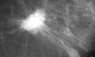
Mammography is a radiographic examination which uses
low-dose radiation to produce high contrast images of
the breast. The purpose of mammography is to detect
breast cancer in the earliest stages of development.
What
is a mammogram?
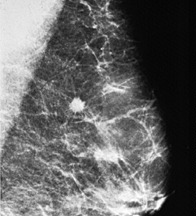
A mammogram is the process of positioning the breast
on a film that will result in a diagnostic image of
the breast tissue.
What
is a mammographer? A mammographer is an individual
trained and skilled in the specialty of mammography.
by
Linda S. Lingar, M.Ed., R.T., (R)(M)
Assistant Professor
Department of Radiologic Technology