|
Lecture 1: |
 |
|
9/21/99
|
Dr. Cooney
|
GeneBiochem
|
Summary Notes
|
L. Van Warren
|
|
Lecture 1: |
|
Section 1: Topics and Readings
Topics
Chromosomes: discrete unit of the genome carrying
many genes.
Half
the mass is protein.
Readings (Lewin - Genes IV)
732-733 Mammalian satellites consist of hierarchical repeats c/o
Chapter 25 Simple Sequence DNA
|
Repetitive DNA is defined by its rapid rate of renaturation. It consists of short sequences that are repeated in identical or related copies in the genome. Highly repetitive DNA consists of very short sequences repeated many times in tandem in large clusters. It is called simple sequence DNA. It is usually less than 10% in mammalian genomes but may be up to 50% in Drosophila virilis. It is often possible to fractionate this due to its distinct buoyant density. This kind of fraction is called satellite DNA. Satellite DNAs are found in regions of heterochromatin. Heterochromatin is tightly coiled up and inert in contrast with the euchromatin that makes up most of the genome. Heterochromatin is frequently found at the centromeres. Arthropods have satellites
with a short repeating unit - only 7 bp. These satellites represent very long stretches of DNA of very low sequence complexity, within which constancy of sequence can be maintained. Mammalian satellites consist of a hierarchy of repeats. |
751 - Loops, Domains and Scaffolds in Eukaryotic DNA
Interphase chromatin is a tangled mass occupying a large part of the nuclear volume, in contrast with the highly organized and reproducible ultrastructure of mitotic chromosomes.
Nuclei can by lysed on top of a sucrose gradient. This releases the genome in a form that can be collected by centrifugation.
Supercoiling occurs about every 200 bp. Supercoils can be removed by nicking with DNAase. Full relaxation requires nicking every 85 kb. These regions could comprise a loop or domain similar to those in bacteria. We want to know whether these loops correspond to specific sequences and whether they have functional significance.
Protein depleted chromosomes take the form of a central scaffold surrounded by a halo of DNA. The appearance of the scaffold takes on the appearance of a mitotic pair of sister chromatids. The sister scaffolds are tightly connected by sometimes separate.
Interphase cells possess a nuclear matrix, a filamentous structure on the interior of the nuclear membrane. Chromatin often appears to be attached to the matrix and it is suggested that this attachment is necessary for transcription or replication.
DNA sites attach to the protein scaffold via MAR (matrix attachment regions) or SAR (scaffold attachment regions).
743 - Chapter 26 Chromosomes
|
6 gigabases fit in the human cell,
and stretches to about 6 feet long.! DNA must be compressed exceedingly tightly to fit into the space available. In contrast with the customary picture of DNA as an extended double helix, structural deformation of DNA to bend or fold it into a more compact form is the rule rather than the exception. In a bacterium the DNA packing
density is 10 mg/ml. Condensing Viral Genomes Into Their Coats
|
762 - The Eukaryotic chromosome as a segregation device
|
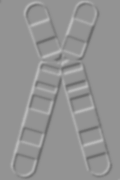
The microtubules comprise a cellular filamentous system, reorganized at mitosis so that they connect the chromosomes to the poles of the cell. The sites in the two regions where the microtubule ends are organized are called MTOC's - microtubule organizing centers. The region of the chromosome that is responsible for its segregation at mitosis and meiosis is called the centromere. It is associated with two important features. It contains the site at which the sister chromatids are held together prior to the separation of the individual chromosomes. This appears as a constricted waist where the four arms of the chromosomes are connected. The term centromere has been used in both the functional and structural sense to describe the feature of the chromosome responsible for its movement. The centromere is pulled towards the pole during mitosis and the attached chromosome is dragged along. Thus the chromosome is MERELY a structure for attaching a large number of genes to the apparatus for division. Quantum thinking baby, yeah! The centromere is essential for segregation as shown by the behavior of chromosomes that have been broken. A single break generates one piece that keeps the centromere - the top of the wishbone as it were - and another, an acentric fragment that lacks it. When translocations generate chromosomes with more than one centromere, aberrant structures form at mitosis, since the two centromeres on the same sister chromatid can be pulled towards different poles, breaking the chromosome. In C-banding centromeres show as darkly stained regions. This kinetochore appears to be directly attached to the microtubles. Fragments of DNA have been isolated by virtue of their ability to confer mitotic stability on these plasmids. A CEN fragment is defined by its ability to confer stability upon such a plasmid. Centromeres are interchangeable. They are used simply to attach the chromosome to the spindle. They play no role in distinguishing one chromosome from another. |
763 - Telomeres seal the ends of chromosomes
|
Telomeres caps the end of DNA. Chromosome ends generated by breakage are "sticky" and tend to react with other chromosomes. It must lie at the end of a chromosome. Cn(A/T)m where n > 1 and m is 1-4. Telomerase is a specialized example
of a reverse transcriptase, an enzyme that synthesizes a DNA sequence
using an RNA template. |
439 -Isolating the origin of yeast replicons
|
Any segment of DNA that has an origin should be able to replicate. Event though plasmids are rare in eukaryotes, it is possible to construct them by suitable manipulation in vitro. This has been accomplished in yeast. S. Cerevisiae mutants can be transformed to the wild phenotype by addition of DNA that carries a wild-type copy of the gene. Some yeast DNA fragments, when circularized, are able to transform defective cells very efficiently. These fragments can survive in the cell as self replicating plasmids. A high-frequency transforming
fragment possesses a sequence that confers the ability to replicate
efficiently in yeast. |
|
Synthesis of Okazaki fragments requires priming, extension, removal of RNA, gap filling, and nick ligation. |
|
A eukaryotic genome is divided into multiple replicons, and the origin in each replicon is activated once and only once in a single division cycle. One round of replication either inactivates or destroys the factor and another round cannot occur until further factor is provided. |
Section 2: Lecture
Topics
Telomeres
--------[TF] -------|---------[]-----[]-------[]--------[]